

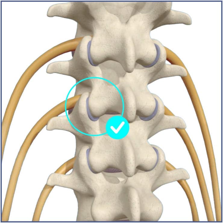
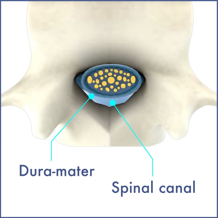
No canal arthrosis, facet capsule with synovial fluid allowing smooth facet joint and spinal motion

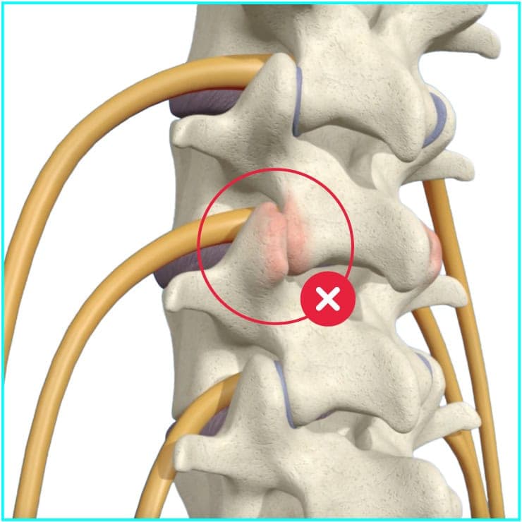
Little fluid left in the facet capsule, the resulting
friction causes inflammation and back pain


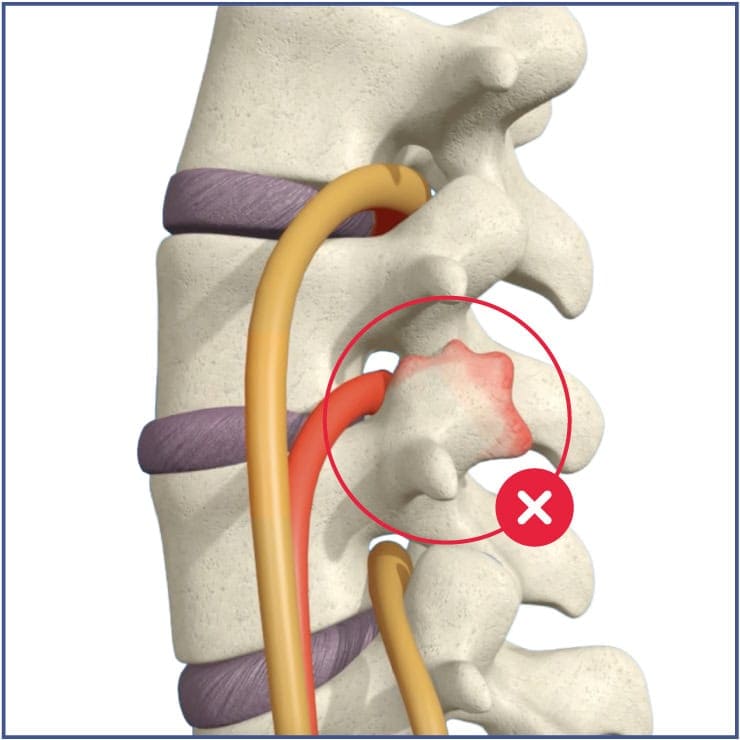
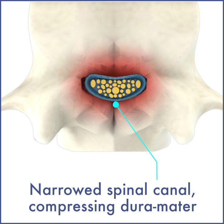
The body naturally responds to this motion/instability by developing bone spurs. Nerve roots are compressed causing additional leg pain and a significantly reduced walking distance. The canal is often narrowed due to arthrosis
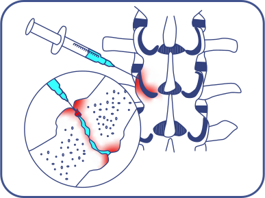
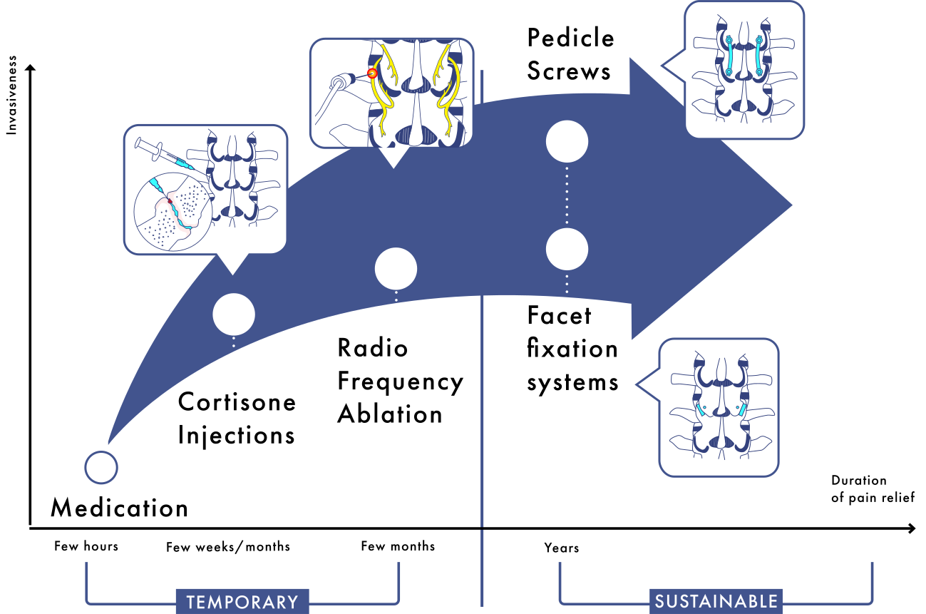
Cortisone injections to reduce facet joint inflammation
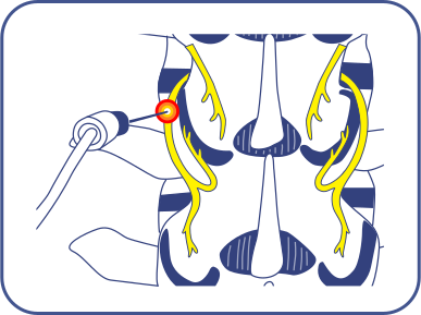
Temporary thermal
destruction of pain nerves
in the facets
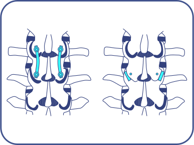
Surgical implantation
of medical devices blocking the facet motion causing pain (pedicle screws, facet fixation systems)
The body naturally responds to this motion/instability by developing bone spurs. Nerve roots are compressed causing additional leg pain and a significantly reduced walking distance. The canal is often narrowed due to arthrosis
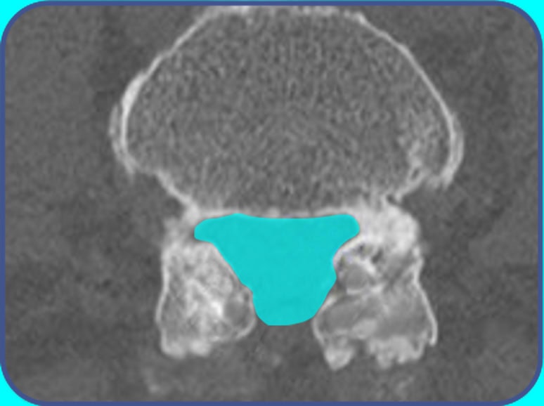
Leg pain is relieved with decompression (bone removal) of canal / foramen. Back pain coming from dura-mater compression is relieved.
If there is back pain coming from the facet joints, it is not addressed by the decompression alone technique.
Decompression alone may also cause postoperative instability and pain over time, potentially requiring a reoperation
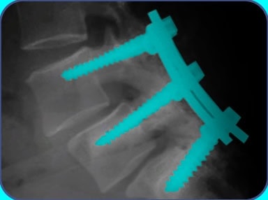
Facet joint back pain and post decompression instability are addressed by adding pedicle screws to the decompression.
Pedicle screw fixation is often required for gross preoperative instability and/or deformity.
The rigidity of the pedicle screw construct may cause levels adjacent to the fixation to degenerate over time, potentially requiring a reoperation.
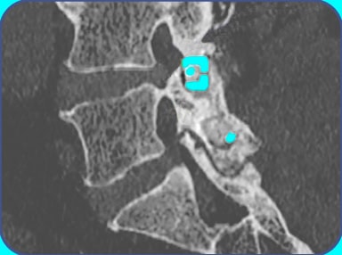
Facet joint back pain and post decompression instability are addressed by adding a facet fixation system to the decompression.
Facet fixation systems can be used in selected patients with up to moderate preoperative instability without deformity.
For these selected patient cases, facet fixation systems can be an alternative to pedicle screws. In two comparative studies in selected patients with lumbar spinal stenosis, facet fixation was associated with shorter surgery and less blood lossSrour, Cureus, 2022 pubmed.ncbi.nlm.nih.gov/35399487/ than pedicle screws, with similar improvement in pain and function at 2 yearsHouari et al., Clin Spine Surg, 2025 (published online 2024) pubmed.ncbi.nlm.nih.gov/39404118/.
Chronic low back pain can come from several structures: the intervertebral discs, the facet joints, the sacroiliac joints, the nerves and the muscles. The facet joints are the source of pain in up to 45%Perolat et al., Insights Imaging, 2018 pubmed.ncbi.nlm.nih.gov/30090998/
Manchikanti et al., Pain Physician, 2008 pubmed.ncbi.nlm.nih.gov/18196171/ of people with chronic low back pain. Facet joint pain is often overlooked, because imaging alone cannot confirm it.
Yes, and they are among its most common sources, involved in up to 45%Perolat et al., Insights Imaging, 2018 pubmed.ncbi.nlm.nih.gov/30090998/
Manchikanti et al., Pain Physician, 2008 pubmed.ncbi.nlm.nih.gov/18196171/ of cases. Wear of the facet joint reduces its lubricating fluid; the resulting friction causes inflammation and pain, typically in the lower back, often worse when standing, twisting or bending backward. These clinical symptoms suggest facet joint pain but cannot confirm it on their own.
Imaging alone cannot confirm that the facet joints are the source of pain, because a significant proportion of people without back pain also show degenerative changes of the facet joints on CT scans or MRIBrinjikji et al., AJNR Am J Neuroradiol, 2015 www.ajnr.org/content/36/4/811. The reference test is a diagnostic block: a local anesthetic is injected into the facet joint or near its small nerves (e.g., medial branch block). If the pain clearly decreases for a few hours, the facet joint is considered the pain generatorCohen et al., Reg Anesth Pain Med, 2020 (multispecialty consensus guidelines) pubmed.ncbi.nlm.nih.gov/32245841/. Your physician interprets the result together with your symptoms, examination and imaging.
Treatment follows steps, from the least to the most invasive. First, conservative care: pain medication, exercise-based physiotherapy and activity adaptation. If pain persists, corticosteroid injections into or around the facet joint, to reduce the painful inflammation locally, with an effect that typically lasts a few weeksCohen et al., Reg Anesth Pain Med, 2020 (multispecialty consensus guidelines) pubmed.ncbi.nlm.nih.gov/32245841/. Then radiofrequency ablation, which uses heat to destroy the small nerves carrying the pain signal to the brain. Relief lasts several months, but the nerves typically grow back after 6 to 12 monthsSmuck et al., PM&R, 2012 (systematic review) onlinelibrary.wiley.com/doi/10.1016/j.pmrj.2012.06.007
Perolat et al., Insights Imaging, 2018 pubmed.ncbi.nlm.nih.gov/30090998/, so the procedure often has to be repeated. When pain keeps returning and the facet joint is confirmed as the source, facet arthrodesis can be discussed in selected patients. The decision is made case by case with a spine physician.
Corticosteroid injections reduce inflammation inside or around the facet joint, but their relief is usually temporary (a few weeks)Perolat et al., Insights Imaging, 2018 pubmed.ncbi.nlm.nih.gov/30090998/
Cohen et al., Reg Anesth Pain Med, 2020 (multispecialty consensus guidelines) pubmed.ncbi.nlm.nih.gov/32245841/. Radiofrequency ablation uses heat to destroy the small nerves that carry the pain signal from the facet joint to the brain. It treats the signal, not its source: the worn joint itself is unchanged. The nerves typically grow back after 6 to 12 monthsSmuck et al., PM&R, 2012 (systematic review) onlinelibrary.wiley.com/doi/10.1016/j.pmrj.2012.06.007
Perolat et al., Insights Imaging, 2018 pubmed.ncbi.nlm.nih.gov/30090998/, so pain returns and the procedure often has to be repeated. When these treatments no longer bring lasting relief, your physician first reassesses the diagnosisCohen et al., Reg Anesth Pain Med, 2020 (multispecialty consensus guidelines) pubmed.ncbi.nlm.nih.gov/32245841/, including other possible causes of pain and spinal instability. When the facet joint has been confirmed as the source, facet arthrodesis can be discussed with your spine physician. Unlike these treatments, it targets the source of the pain: it is intended to fuse the painful facet joints, so that the abnormal motion (micro-motion) that causes the inflammationVan Oosterwyck et al., Life, 2024 www.mdpi.com/2075-1729/14/11/1480 stops.
Facet arthrodesis (facet fusion) is a surgical procedure intended to fuse the painful facet joints so that they no longer move and rub. A facet joint is a small joint at the back of the spine, between two vertebrae. When it wears out, it moves abnormally with every step and every bending movement. These small abnormal movements (micro-motion) stretch the joint capsule and irritate the joint lining, which causes painful inflammationVan Oosterwyck et al., Life, 2024 www.mdpi.com/2075-1729/14/11/1480. Fusion means making two bones grow together into one solid bone. Once the joint is fused, it can no longer move, so the micro-motion and the inflammation it causes stop. To achieve this, the surgeon prepares the joint surfaces, adds bone graft and places a small implant that holds the joint still to promote new bone growth across it, much like a cast holds a broken bone to help it heal. This implant can be a facet cage (spacer) placed inside the joint, a screw across it, or both. It can be performed through open surgery or through minimally invasive approaches (tubular, endoscopic or fully percutaneous). Unlike conventional fusion with Pedicle screws and rods, it acts only on the facet joints. Fusion develops progressively: in published studies of facet arthrodesis with facet screws or facet cages, it was confirmed on imaging about one year after surgery in 86% to 100% of casesMusso et al., J Craniovertebr Junction Spine, 2022 (systematic review, 19 studies, 1,577 patients) pmc.ncbi.nlm.nih.gov/articles/PMC9910129/.
Conventional spinal fusion is intended to join two or more vertebrae so that they fuse into a single solid block of bone. It usually uses Pedicle screws, inserted into each vertebra and connected by rods, often with a cage placed in the disc space after removal of the disc. Whether there is an alternative depends on the cause of the pain, the stability of the spine and the patient's anatomy. Some options avoid fusion altogether, such as decompression alone in lumbar spinal stenosis, when the spine is stable and there is no significant facet joint pain. Others keep fusion but make it lighter: facet arthrodesis is intended to fuse only the facet joints of the painful segment, without Pedicle screws, rods or removal of the disc. It is a lighter form of fusion rather than an alternative to fusion, and it is not suitable for every patient: major instability or deformity still requires Pedicle screws.
Lumbar spinal stenosis is a narrowing of the spinal canal or of the openings through which the nerves exit, in the lower back. With age, worn facet joints enlarge, ligaments thicken and discs bulge, and together they squeeze the nerves. The typical symptom is pain, heaviness or numbness in the legs when walking or standing, relieved by sitting or bending forward. Many patients also have low back pain, which can come from the worn facet jointsPerolat et al., Insights Imaging, 2018 pubmed.ncbi.nlm.nih.gov/30090998/. When conservative care is not enough, surgery frees the nerves by removing what compresses them (decompression). Decompression alone treats the nerve compression, but in a registry of more than 25,000 patients, only about half reported a substantial reduction of their back painKnight et al., J Neurosurg Spine, 2023 (British Spine Registry, 25,349 patients) pubmed.ncbi.nlm.nih.gov/37486861/ (by half or more). Since the nerves have been freed and the dura mater (the membrane surrounding them) decompressed, the facet joints are one of the main remaining sources of this back pain. When facet joint pain is present, or when spinal instability is present or expected after the decompression, a fusion of the affected segment can be performed during the same operation.
The choice depends mainly on the stability and alignment of the spine. In selected patients without major instability or deformity, facet arthrodesis can be used instead of Pedicle screws, for example when a fusion is added to a decompression for lumbar spinal stenosis. Pedicle screws remain necessary for major instability or deformity, or when the alignment of the spine must be corrected. Talk to your spine physician about which technique suits your case.
Where do you live?